
Image Viewer
An image viewer displays the microscopy image data contained in a SPARC dataset.
Image Viewer
An image viewer displays the microscopy image data contained in a dataset.
Control the image viewer as follows:
-
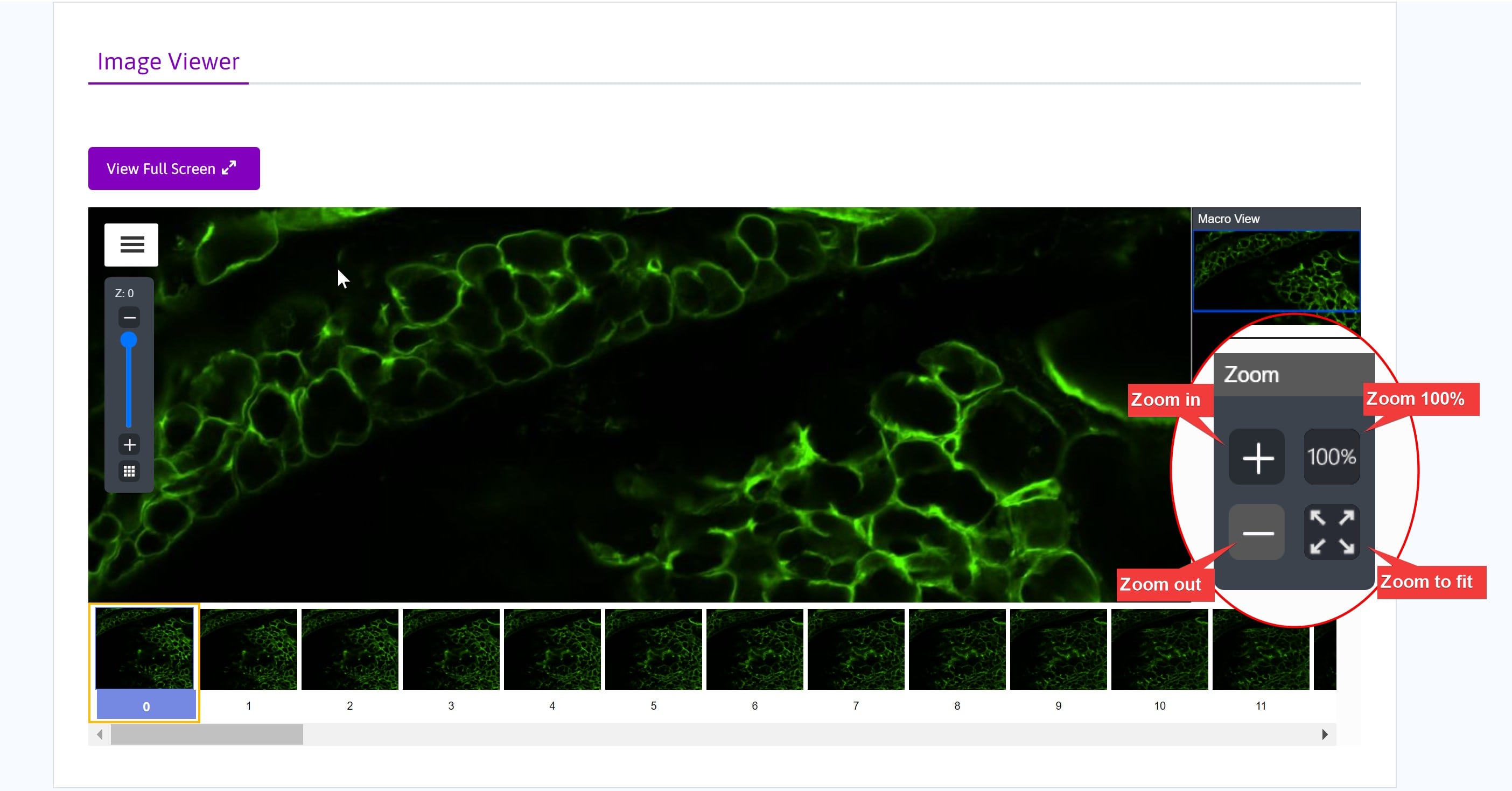
Zoom in and zoom out of the image by:
- scrolling with the mouse wheel or
- clicking the (+) and (-) buttons in zoom controls
-
Additional zoom controls:
-
Click the 100% button to jump to Zoom 100%
-
Fit the image to the window using the Zoom to Fit button
-
The dynamic scale bar updates as you zoom in and out of the image.
-
The Macro View window displays a mini view of the entire image and outlines the region displayed in the main viewer window. Click in the macro view window to display that portion of the image.

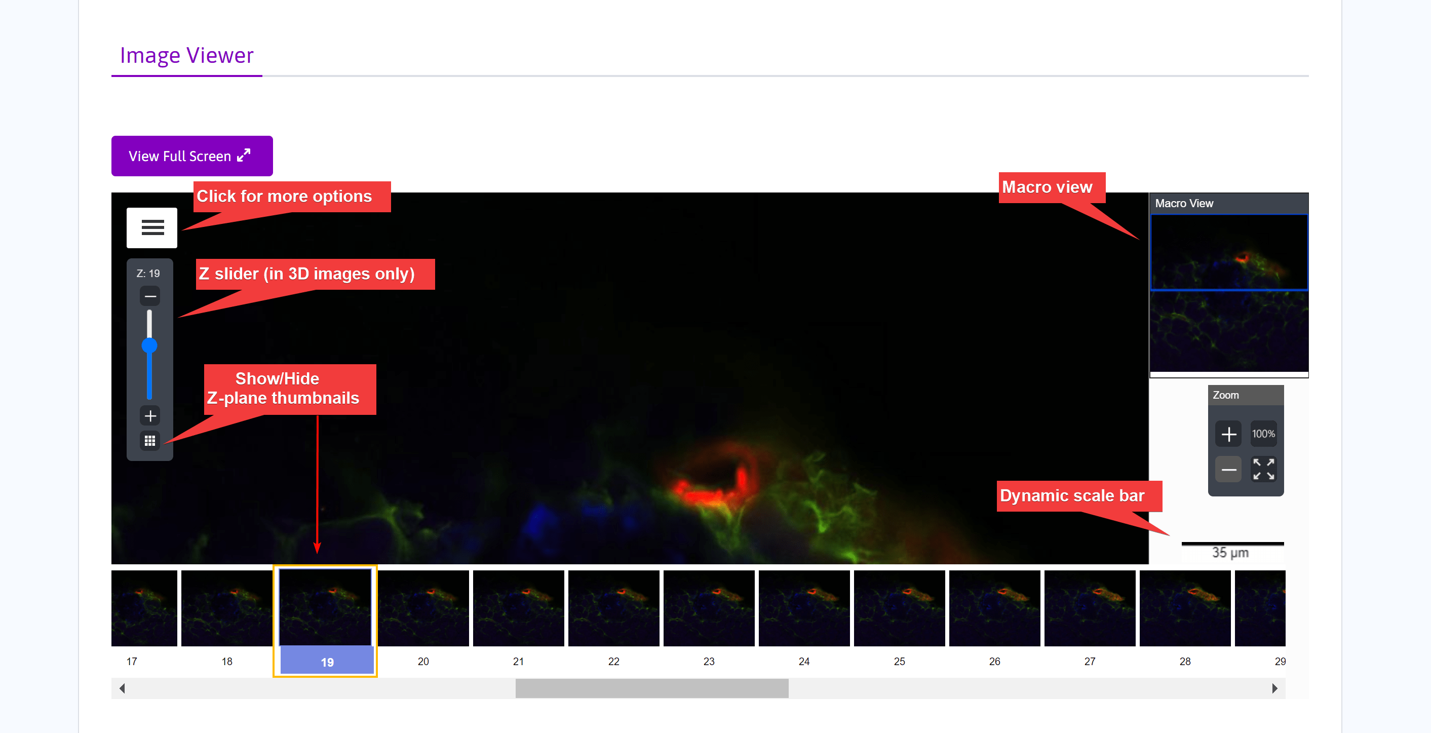
- The Z slider is displayed if the image is 3D. Move through the image Z planes by dragging the slider. Use the up- and down-arrow buttons to move through the image Z planes one at a time.
- Click View focal image thumbnails to toggle the display of thumbnail images for each Z plane below the main viewer window. Use the scroll bar or the (+) and (-) buttons to move through the Z plane thumbnails. Click a thumbnail image to select and view that Z position.
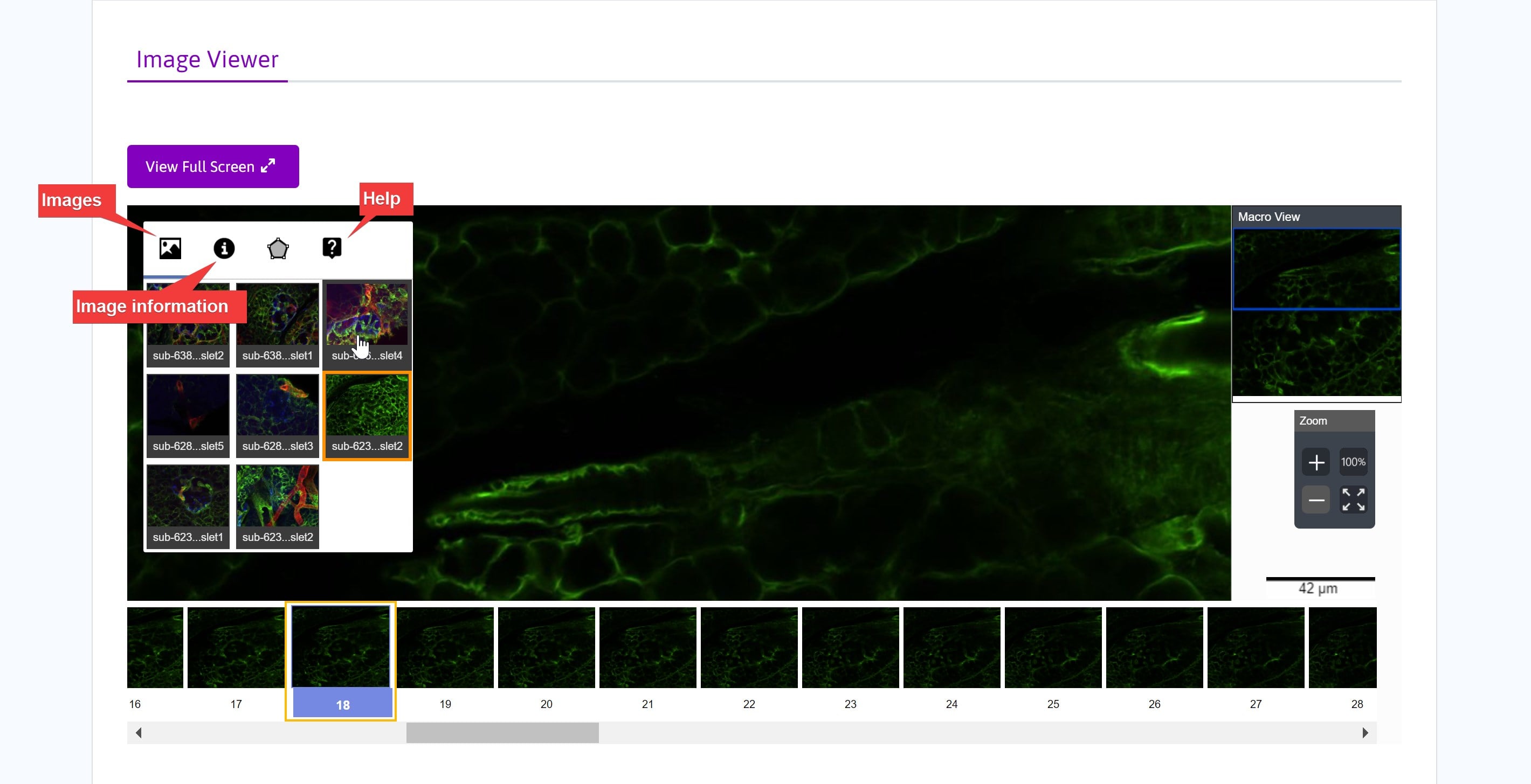
- Select More information to reveal more options in the Image Viewer.
- Click Images to view thumbnails of other images in the dataset collection. Clicking a thumbnail will launch the Biolucida Web Viewer in another window and display the image.
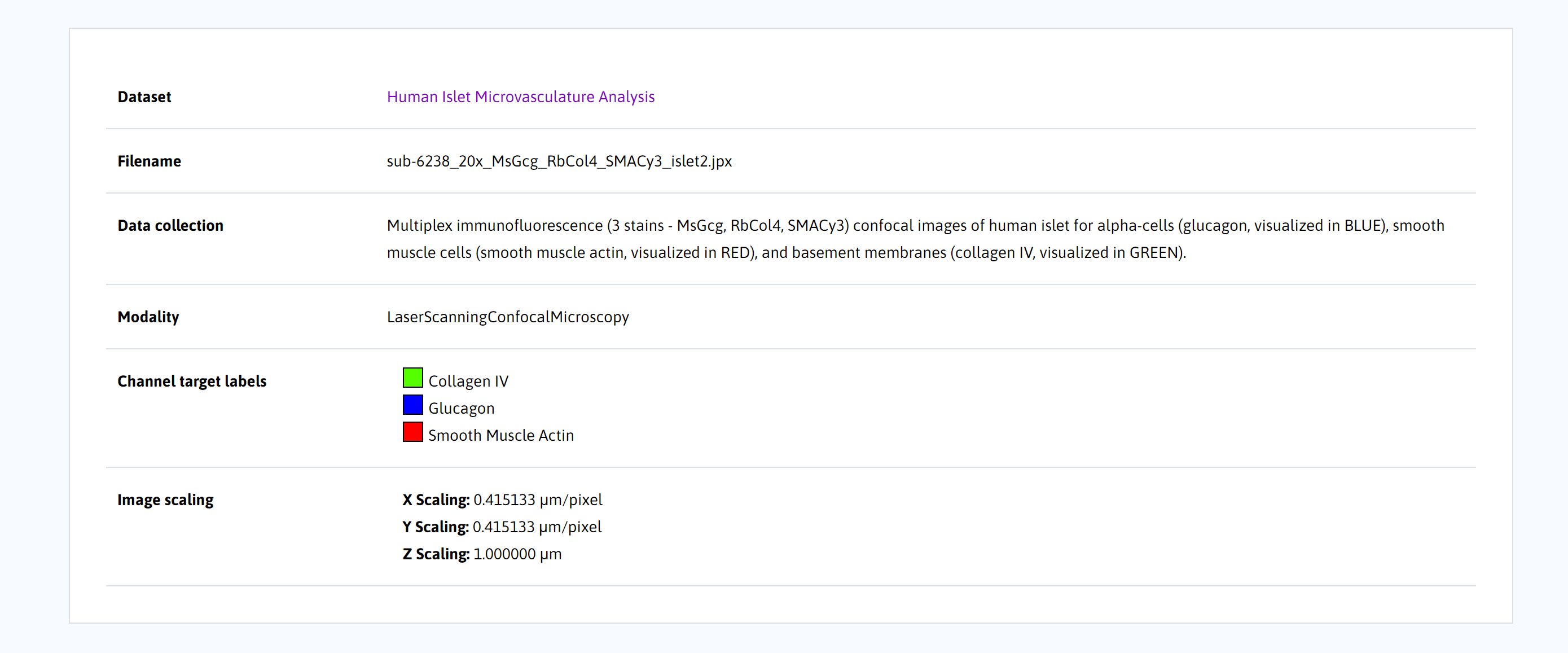
- Click Image Information to view information about the current image.
- Clicking Help will take you to the Biolucida User Guide.
- Below the image, file-level metadata is provided, including modality, channel target labels, and image scaling.
Updated almost 2 years ago